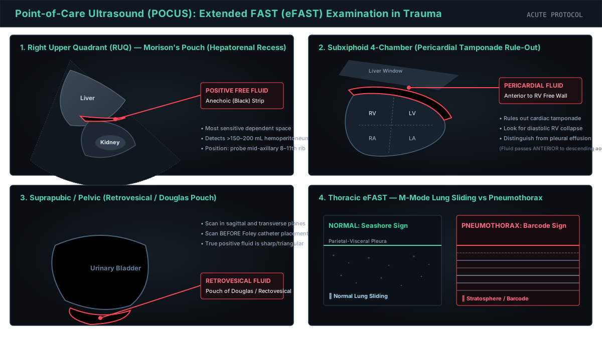
Four-panel Point-of-Care Ultrasound (POCUS) eFAST trauma diagnostic schematic: (A) Right Upper Quadrant Morison pouch showing anechoic free fluid stripe between liver and right kidney; (B) Subxiphoid 4-chamber cardiac view showing circumferential pericardial effusion compressing the right ventricle; (C) Pelvic suprapubic sagittal view demonstrating hemoperitoneum pooling in the pouch of Douglas posterior to the bladder; (D) Thoracic pleural assessment comparing normal M-mode Seashore sign (granular lung sliding) with pneumothorax Barcode / Stratosphere sign.
“Point-of-Care Ultrasound: eFAST Trauma Diagnostic Protocol” by ClinInterpret Medical Education Editorial Board — CC BY 4.0. View Full Image.
Common Pitfalls & Diagnostic Traps
- Confusing the physiological fluid in the gallbladder or inferior vena cava for pathological free fluid in Morisons pouch; ensure the probe is angled to identify the distinct echogenic renal capsule and liver parenchyma.
- Relying on FAST to rule out retroperitoneal bleeding or visceral organ injury in hemodynamically stable trauma patients; FAST has high specificity (>95%) but only moderate sensitivity (65-80%) for blunt injury without hemoperitoneum.
- Mistaking an epicardial fat pad for a pericardial effusion on the subxiphoid view; fat pads are typically anterior, hypoechoic with low-level internal echoes, and move synchronously with the ventricular myocardium, whereas pericardial effusion is jet-black (anechoic) and accumulates dependently behind the posterior LV wall.
- Misinterpreting the 'Stratosphere Sign' caused by a patient breath-hold or apnea as a pneumothorax; verify the absence of B-lines and search for a definitive Lung Point before diagnosing pneumothorax in an unventilated patient.
1Overview and Physiology
Point-of-Care Ultrasound (POCUS) via the Focused Assessment with Sonography for Trauma (FAST) and Extended FAST (eFAST) protocol is the standard-of-care rapid bedside imaging modality performed during the primary survey of trauma resuscitations. Its primary clinical mandate is to identify life-threatening hemorrhage and air accumulation within non-expandable physiological spaces: the pericardium, peritoneal cavity, and pleural spaces.
2Ultrasound Physics & Acoustic Properties in Trauma
- Piezoelectric Transduction & Frequency:
- Diagnostic ultrasound transmits high-frequency acoustic waves (1 to 15 MHz) generated by piezoelectric crystals.
- Low-frequency curvilinear transducers (2 to 5 MHz) provide deep tissue penetration (15 to 30 cm) suitable for transabdominal Morrison's pouch and pelvic interrogation.
- Phased array transducers (1 to 5 MHz) utilize a small footprint designed to fit between narrow intercostal spaces for subxiphoid and parasternal cardiac imaging.
- High-frequency linear transducers (7 to 12 MHz) provide high spatial resolution for superficial structures (1 to 5 cm), optimal for the pleural interface and pneumothorax detection.
- Acoustic Impedance & Echogenicity:
- Biological tissues resist sound propagation based on density (rho) and acoustic velocity (c): Z = rho × c.
- Anechoic (Jet-Black): Pure fluids (blood, serous fluid, urine, transudate) transmit acoustic waves without internal reflection. Free hemoperitoneum appears as sharp, conforming black fluid wedges.
- Hyperechoic (Bright White): Connective tissue, fibrous organ capsules (Glisson's capsule, Gerota's fascia), bone cortices, and tissue-gas boundaries strongly reflect sound waves.
- Hypoechoic (Medium to Dark Grey): Solid organ parenchyma (liver, spleen, renal cortex, myocardium) produces intermediate acoustic backscatter.
- Acoustic Enhancement: Fluid collections transmit sound with minimal attenuation, causing deep parenchymal tissues immediately posterior to fluid to appear brighter (posterior acoustic enhancement).
3Technique and Quality Checks
- Transducer Selection and Frequency Matching:
- Use a low-frequency curvilinear probe (2--5 MHz) for abdominal, pelvic, and pleural fluid assessment.
- Use a phased-array cardiac probe (1--5 MHz) for subxiphoid or parasternal pericardial assessment.
- Use a high-frequency linear vascular probe (7--12 MHz) for anterior thoracic pleural sliding and pneumothorax detection.
- Preset and Gain Optimization:
- Select the dedicated 'Abdomen' or 'Trauma' preset to minimize post-processing smoothing and optimize contrast between echogenic capsule borders and anechoic free fluid.
- Adjust overall gain so that the interior of normal blood vessels (IVC, aorta) and full bladder appear uniformly anechoic (pure black), while liver and renal parenchyma display mid-level homogeneous grey echoes.
- Depth and Sector Focus:
- Set the initial depth to 15--20 cm in the RUQ and LUQ views to ensure visualization of the posterior acoustic boundary (diaphragm and retroperitoneum). Shallow depth settings risk missing dependent retrohepatic or retrorenal fluid stripes.
- Position the focal zone directly at the depth of the hepatorenal and splenorenal interfaces.
- Respiration and Probe Sweep:
- Complete assessment requires multi-planar, three-dimensional sweeping (fanning) through each anatomical window from anterior to posterior margins, not static snapshots.
- In spontaneously breathing patients, evaluate fluid redistribution across the respiratory cycle.
4Normal Anatomy
Systematic appraisal begins with recognition of baseline normal acoustic landmarks across the standard FAST windows:
- Right Upper Quadrant (RUQ / Morison's Pouch):
- Homogeneous granular echotexture of the liver parenchyma (left of screen).
- Normal corticomedullary differentiation of the right kidney with echogenic renal capsule and sinus fat.
- Potential space between Glisson's capsule and Gerota's fascia shows zero fluid separation.
- Echogenic curvilinear diaphragmatic line separating abdominal contents from the air-filled right lung base.
- Left Upper Quadrant (LUQ / Splenorenal Recess):
- Finely granular splenic parenchyma abutting the left hemidiaphragm superiorly.
- Left kidney oriented slightly more posterior and superior than the right kidney.
- Intact splenodiaphragmatic, splenorenal, and retroperitoneal spaces without acoustic disruption.
- Pelvic / Suprapubic Space:
- Distended urine-filled urinary bladder serving as an acoustic window with thin, smooth hyperechoic muscular walls.
- In males: Well-defined rectovesical space with prostate and seminal vesicles.
- In females: Uterus positioned posterior to bladder, separating vesicouterine and rectouterine (pouch of Douglas) potential spaces.
- Subxiphoid Pericardial Space:
- Right ventricular free wall directly abutting the left hepatic lobe acoustic window.
- Normal physiological pericardial sliding (shimmering hyperechoic visceral and parietal pericardial layers).
- Absence of fluid separation between epicardium and parietal pericardium.
- Thoracic Pleural Interface:
- Bright, glistening hyperechoic pleural line approximately 0.5--1.0 cm deep to the rib interfaces.
- Normal visceral-on-parietal pleural sliding with tidal ventilation.
- Horizontal reverberation A-lines and occasional vertical comet-tail B-lines.
5Key Signs and Patterns
$begin{array}{|l|l|l|} hline textbf{Sonographic Sign} & textbf{Physical / Acoustic Appearance} & textbf{Pathological Meaning} \ hline Anechoic Stripe & Conforming jet-black fluid stripe between tissue planes & Free hemoperitoneum / hemothorax / effusion \ Seashore Sign & Motionless stripes above pleural line + granular sand below (M-mode) & Normal aerated lung with intact visceral pleural sliding \ Stratosphere / Barcode & Uniform horizontal parallel lines across all depths (M-mode) & Pneumothorax (absence of pleural sliding) \ Lung Point (Point d'Appel) & Respiratory alternating Seashore and Barcode patterns at margin & Pathognomonic boundary of pneumothorax (100% specific) \ Spine Sign & Continuation of thoracic vertebral acoustic reflections above diaphragm & Pleural effusion or hemothorax (fluid transmits acoustic wave) \ Acoustic Mirror Artifact & Duplicate liver/spleen parenchyma visible above diaphragm & Normal aerated lung base (confirms absence of hemothorax) \ hline end{array}$
6Indications
- Blunt Abdominal and Torso Trauma: High-speed motor vehicle crashes, pedestrian vs automobile, falls from height, crush injuries, and direct blows to flank or abdomen.
- Penetrating Torso Trauma: Stab and gunshot wounds to the 'trauma box' (clavicles to groin, anterior and posterior), specifically assessing for occult hemopericardium, cardiac tamponade, and pneumothorax.
- Undifferentiated Hypotension and Shock in Trauma: Rapid identification of internal exsanguinating hemorrhage vs obstructive shock (tension pneumothorax, tamponade).
- Acute Thoracic Trauma: Rapid evaluation for traumatic pneumothorax and massive hemothorax during primary survey without waiting for portable radiographs.
- Procedural Guidance: Real-time ultrasound localization for emergent tube thoracostomy, pericardiocentesis, or diagnostic peritoneal aspiration.
7Systematic Interpretation Steps
Step 1: Subxiphoid Pericardial Window
   ├─ Flat probe against epigastrium, angled toward left shoulder using liver acoustic window
   ├─ Inspect for anechoic pericardial fluid stripe separating epicardium from parietal pericardium
   └─ Assess for right ventricular diastolic collapse (diagnostic of cardiac tamponade)

Step 2: Right Upper Quadrant (Morison's Pouch) Sweep
   ├─ Mid-axillary line 8th-11th ICS; sweep through entire 3D volume of Morison's pouch
   ├─ Inspect inferior hepatic tip, hepatorenal interface, and subdiaphragmatic space
   └─ Slide cephalad across diaphragm to check for hemothorax and positive spine sign

Step 3: Left Upper Quadrant (Splenorenal) Sweep
   ├─ Posterior axillary line 6th-9th ICS ('knuckles on stretcher'); sweep spleen and diaphragm
   ├─ Inspect subdiaphragmatic space (primary fluid site) and splenorenal interface
   └─ Slide cephalad to evaluate left pleural space and supradiaphragmatic spine sign

Step 4: Pelvic Suprapubic Sweep
   ├─ Transverse probe superior to pubic symphysis, angled inferiorly into true pelvis
   ├─ Rotate 90 degrees to sagittal orientation; sweep entire rectovesical / Douglas recess
   └─ Check for free anechoic fluid pooling behind the posterior bladder wall

Step 5: Extended Thoracic Pleural Windows
   ├─ Switch to high-frequency linear transducer (7-12 MHz) in lung preset
   ├─ Interrogate 2nd to 4th ICS at mid-clavicular line bilaterally
   ├─ Confirm pleural sliding and presence of B-lines on B-mode
└─ Position M-mode cursor over pleural line to confirm Seashore Sign vs Barcode Sign8Abnormal Patterns and Differential Diagnosis
91. Hemoperitoneum (Positive FAST Abdomen)
- Sonographic Findings: Sharp, wedge-shaped or ribbon-like anechoic (jet-black) fluid collections separating organ capsules in Morison's pouch, the splenorenal recess, or the rectovesical/Douglas pouch.
- Differential Diagnosis: Hemoperitoneum (trauma), pre-existing ascites (cirrhosis, peritoneal dialysis, heart failure), ruptured ectopic pregnancy, ruptured ovarian cyst, or ruptured hollow viscus peritoneal fluid.
102. Hemopericardium and Cardiac Tamponade (Positive FAST Pericardium)
- Sonographic Findings: Anechoic fluid rim tracking between the epicardium and parietal pericardium. In hemodynamic tamponade, early diastolic inward buckling (collapse) of the thin-walled right ventricle and late diastolic collapse of the right atrium are diagnostic.
- Differential Diagnosis: Traumatic hemopericardium, chronic pericardial effusion (malignancy, uremia, tuberculosis), anterior epicardial fat pad (hypoechoic, moves with heart, lacks posterior fluid).
113. Traumatic Pneumothorax (Positive eFAST Thorax)
- Sonographic Findings: Complete absence of B-mode pleural sliding, absence of comet-tail B-lines, replacement of Seashore sign with horizontal Stratosphere / Barcode sign on M-mode, and discovery of a pathognomonic Lung Point.
- Differential Diagnosis: Traumatic pneumothorax, severe bullous emphysema, right mainstem bronchus intubation (contralateral lung immobility), pleurodesis/pleural symphysis, apnea/respiratory arrest.
12Limitations and Pitfalls
- Fluid Volume Threshold: FAST requires 200--400 mL of free peritoneal blood for reliable acoustic detection. A negative FAST in early resuscitation does NOT exclude ongoing low-volume intra-abdominal bleeding.
- Blind Spots: FAST does not visualize the retroperitoneum (pancreas, aorta, duodenal trauma, retroperitoneal hematomas from pelvic ring fractures).
- Parenchymal Organ Injury: FAST detects free fluid, not solid organ tears. Contained subcapsular hematomas or parenchymal lacerations without peritoneal leakage will yield a 'false-negative' FAST.
- Empty Bladder Pitfall: A catheterized or decompressed urinary bladder eliminates the acoustic transmission window for pelvic fluid interrogation.
- Body Habitus and Subcutaneous Air: Severe obesity, intestinal bowel gas, and extensive subcutaneous emphysema degrade beam transmission and acoustic resolution.
13Critical Findings and Red Flags
> [!CAUTION] > Emergency Surgical Escalation Triggers: > 1. Hemodynamically Unstable Patient with Positive Abdominal FAST: Transfer immediately to the operating theater for emergent exploratory laparotomy. Under no circumstances should an unstable trauma patient undergo CT scanning! > 2. Pericardial Fluid with Right Ventricular Diastolic Collapse: Emergency resuscitative subxiphoid pericardial window, bedside needle pericardiocentesis, or resuscitative thoracotomy. > 3. Absent Lung Sliding with Barcode Sign and Hemodynamic Shock: Presumptive tension pneumothorax; perform immediate needle decompression or finger thoracostomy before awaiting chest radiography.
14Exam Pearls
- The Order of Fluid Accumulation: In the RUQ, fluid gathers first at the inferior tip of the right liver lobe, not in the deep hepatorenal crevice. In the LUQ, fluid gathers first between the spleen and the diaphragm, not the splenorenal recess.
- The Power of the Lung Point: Finding a Lung Point is 100% specific for pneumothorax and allows estimation of pneumothorax size by how far posteriorly the lung point is displaced along the chest wall.
- Acoustic Spine Sign: On a normal scan, sound waves are reflected by aerated lung, casting a dark acoustic shadow where the thoracic spine cannot be seen above the diaphragm. When hemothorax or pleural effusion is present, the fluid conducts sound, revealing bright vertebral bodies marching cephalad past the diaphragm.
- ATLS Protocol Mandate: FAST is a rule-in test for emergency operative intervention in hemodynamically unstable patients; in stable patients, contrast-enhanced CT of the abdomen and pelvis remains the gold standard.
Related Investigations & Companion Tests
Attributed Textbooks & Clinical Guidelines
- American College of Emergency Physicians (ACEP). Ultrasound Guidelines: Emergency, Point-of-Care, and Clinical Ultrasound in Medicine. Ann Emerg Med. 2017;69(5):e27-e54
- American College of Surgeons (ACS) Committee on Trauma. Advanced Trauma Life Support (ATLS) Student Course Manual. 10th ed. 2018
- Kirkpatrick AW et al. The FAST, and eFAST, ultrasound for trauma: A review. J Trauma Acute Care Surg. 2014;76(2):495-502
- Lichtenstein DA. BLUE-protocol and FALLS-protocol: two applications of lung ultrasound in the critically ill. Chest. 2015;147(6):1659-1670
What are the four traditional acoustic anatomical windows of the standard FAST exam, and what two views are added in the extended FAST (eFAST)?
Notes & Highlights
Saved locally onlyA 28-year-old male is brought to the emergency department following a high-speed rollover motor vehicle collision. On arrival, he is pale, diaphoresis is noted, blood pressure is 78/42 mmHg, heart rate is 134 bpm, and respiratory rate is 28/min. A bedside FAST examination is performed during primary survey resuscitation. An anechoic (jet-black) fluid stripe measuring 1.8 cm is visualized separating the liver from the superior pole of the right kidney in the hepatorenal recess. The subxiphoid cardiac view reveals no pericardial fluid. What is the immediate, definitive management step?
A 35-year-old unrestrained driver with severe blunt chest trauma presents with worsening respiratory distress and hypoxemia. Bedside extended FAST (eFAST) of the right hemithorax using a high-frequency linear transducer placed at the 3rd intercostal space mid-clavicular line shows complete absence of pleural sliding (motionless shimmering pleural line) and absence of vertical comet-tail B-lines. When M-mode is placed across the pleural line, a 'barcode' (stratosphere) pattern is seen. Scanning laterally reveals a focal boundary where normal lung sliding and seashore pattern intermittently appear and disappear synchronously with tidal respiration. What does this transition boundary represent, and what is its clinical significance?
A 42-year-old pedestrian struck by an SUV is admitted to the trauma resuscitation bay with pelvic pain. GCS is 15, BP is 128/82 mmHg, HR is 88 bpm. Bedside FAST examination is performed by an emergency physician: RUQ, LUQ, pelvic, and subxiphoid views reveal no anechoic free fluid. The patient remains hemodynamically stable. What is the most appropriate interpretation and next clinical action?