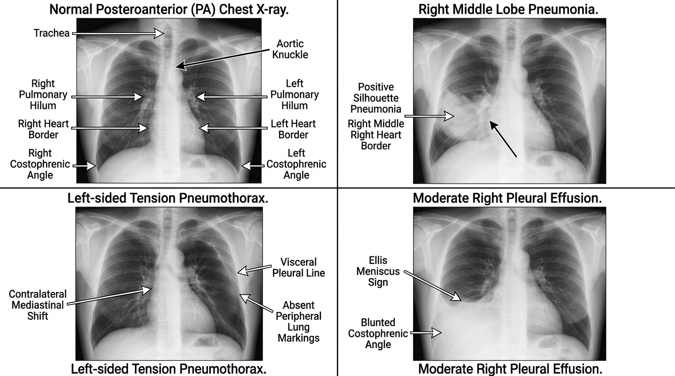
Four-panel clinical chest radiograph diagnostic comparison: (A) Normal PA projection with anatomical ABCDE review and sharp costophrenic angles; (B) Right middle lobe consolidation demonstrating positive silhouette sign obscuring the right heart border; (C) Tension pneumothorax showing marked right lung collapse, visceral pleural line, and contralateral tracheal/mediastinal shift; (D) Right pleural effusion demonstrating dense basilar opacification with classic lateral Ellis meniscus sign.
“Chest Radiograph Multi-Panel Diagnostic Suite” by ClinInterpret Medical Education Editorial Board — CC BY 4.0. View Full Image.
Common Pitfalls & Diagnostic Traps
- Diagnosing cardiomegaly on an Anteroposterior (AP) chest radiograph; the divergence of X-ray beams magnifies the anterior cardiac silhouette by 15-20% on AP projections. Cardiothoracic Ratio (> 0.50) is valid ONLY on an erect Posteroanterior (PA) radiograph.
- Calling a lower zone opacity 'consolidation' when it is actually basilar atelectasis from inadequate inspiratory effort; always count posterior ribs (minimum 9-10 visible) before diagnosing lower lobe opacification in hypoventilating patients.
- Missing pneumothoraces on supine trauma or ICU films because of expecting an apical crescent; on supine radiographs, look for the Deep Sulcus Sign (lucency extending into the anterior/lateral costophrenic angle) and hyperlucent cardiac contours.
- Overlooking the five classic CXR 'hidden areas': apices (apical lung cancer / Pancoast tumor), retrocardiac space (left lower lobe consolidation or hiatus hernia), retrosternal space, below the diaphragm (subdiaphragmatic free air), and the bilateral hila.
1Overview and Physiology
The plain Chest Radiograph (CXR) remains the single most frequently performed diagnostic imaging examination in clinical medicine. It utilizes ionizing electromagnetic radiation (photons) emitted from a cathode-anode X-ray tube, directed through the thoracic cavity, and captured on a digital detector plate or phosphor imaging receptor.
2Physics of Radiodensity and Attenuation
The diagnostic utility of plain radiography is rooted in the differential attenuation of X-ray photons by biological tissues of varying physical densities and atomic numbers (Z). In clinical radiography, biological materials are grouped into five fundamental radiological densities (from most radiolucent / black to most radiopaque / white):
$begin{array}{|l|l|l|} hline textbf{Radiological Density} & textbf{Physical Nature} & textbf{Radiological Appearance} \ hline Air / Gas & Alveoli, trachea, bronchi, gastric bubble & Uniformly Radiolucent (Black) \ Fat & Epicardial fat pad, subcutaneous tissue, fascial planes & Dark Grey \ Soft Tissue / Fluid & Heart, great vessels, blood, blood cells, exudate, transudate & Intermediate / Light Grey \ Bone / Mineral & Ribs, clavicles, scapulae, spine, calcified granulomas & Radiopaque (Off-White) \ Metal / Contrast & Prosthetic valves, pacemaker leads, surgical clips, barium & Radiodense (Bright White) \ hline end{array}$
Because soft tissue (myocardium, pericardial fluid) and water/fluid (pleural effusion, alveolar transudate, pus) possess virtually identical linear attenuation coefficients, they display the exact same radiological shade of grey. A structure is visualized on a plain CXR only when its boundary is outlined by an interface with a material of different physical radiodensity (most typically air in the aerated lung parenchyma).
3Technique and Quality Checks
Before interpreting any thoracic pathology, the clinician must execute a rigorous four-point technical assessment using the mnemonic R-I-P-E (Rotation, Inspiration, Penetration, Exposure / Projection).
41. Rotation
- Assessment Method: Identify the medial heads of both clavicles and the central spinous processes of the upper thoracic vertebrae. The distance from the right clavicular head to the spinous process must equal the distance from the left clavicular head to the spinous process.
- Clinical Consequence: Patient rotation toward the left causes the right clavicle to appear further from the spinous process and falsely projects the mediastinal and hilar shadows toward the right, mimicking mediastinal widening or lung distortion.
52. Inspiration (Lung Volume)
- Assessment Method: Count the number of visible ribs above the dome of the right hemidiaphragm at the mid-clavicular line.
- Posterior ribs run horizontally and are easily identified by their direct origin from the vertebral bodies.
- Anterior ribs angle downward and medially at roughly a 45-degree angle.
- Adequacy Standard: A fully inspiratory radiograph demonstrates 9 to 10 visible posterior ribs (or 6 anterior ribs).
- Clinical Consequence: Poor inspiratory effort (hypoinspiration) bunches normal pulmonary vascular markings at the lung bases, mimicking bibasilar alveolar consolidation or pulmonary edema, while elevating the diaphragm and falsely expanding the cardiothoracic ratio.
63. Penetration (X-Ray Energy / Beam Hardness)
- Adequacy Standard: The lower thoracic vertebral bodies and their intervening intervertebral disc spaces should be just faintly identifiable through the central cardiac silhouette. Pulmonary retrocardiac vessels should be discernible.
- Underpenetrated Film (Too Light): Thoracic spine is invisible behind the heart. Normal lung parenchyma appears falsely hazy or infiltrated; mild infiltrates are exaggerated.
- Overpenetrated Film (Too Dark): The lungs appear uniformly black; subtle nodular opacities, fine reticular interstitial markings, and early ground-glass infiltrates are burned out and erased.
74. Exposure and Projection: PA vs. AP
- Posteroanterior (PA) Projection (Standard Erect):
- The patient stands upright facing the detector cassette; X-ray beams travel from posterior to anterior (1.8 meters / 6 feet tube-to-detector distance).
- The heart, situated anteriorly in the chest, sits directly against the film detector. Geometric magnification is negligible (< 5%).
- Scapulae are rolled forward and rotated away from the lung fields.
- Anteroposterior (AP) Projection (Bedside / Portable):
- The X-ray cassette is placed behind the supine or semi-erect patient's back; beams travel anterior to posterior (1.0 meter distance).
- The anterior heart is far from the detector plate, causing significant divergent beam magnification (15% to 20% magnification of cardiac width).
- Scapulae fall within the mid- and upper lung zones, obscuring parenchymal details.
- Pulmonary blood flow redistributes to the upper lobes simply due to recumbency, mimicking pathological venous cephalization.
8Normal Anatomy
91. Mediastinal and Cardiac Contours
- Right Mediastinal Border: Formed superiorly by the vertical border of the Superior Vena Cava (SVC) and inferiorly by the gently convex curve of the Right Atrium (RA). The Right Ventricle is an anterior structure and does NOT form any border on a normal PA radiograph.
- Left Mediastinal Border: Formed superior-to-inferior by:
- Aortic Knuckle (Knob): The transverse aortic arch curving posteriorly.
- Aortopulmonary Window: Concave space between aortic knob and left pulmonary artery; obliteration suggests mediastinal lymphadenopathy.
- Main Pulmonary Artery Trunk (Conus): Mild straight or gently concave contour.
- Left Atrial Appendage: Barely visible indentation; becomes prominent when the left atrium enlarges.
- Left Ventricle (LV): Broad, convex lateral sweep tapering into the cardiac apex.
102. Trachea and Bronchial Tree
- The trachea is normally midline or slightly shifted to the right at the level of the aortic arch.
- The Carina bifurcates into the right mainstem bronchus (more vertical, wider, 20–30^° angle) and left mainstem bronchus (more horizontal, 40–50^° angle). Normal carinal subcarinal angle is < 90^°; widening indicates subcarinal lymphadenopathy or left atrial dilation.
113. Diaphragm and Pleural Recesses
- The Right Hemidiaphragm sits approximately 1.0 to 2.5 cm higher than the Left Hemidiaphragm due to hepatic mass beneath it and downward displacement by the cardiac apex on the left.
- Costophrenic Angles: Sharp, acute, deep wedges formed by the chest wall and the diaphragm.
- In an upright adult, $150\text{–}200\ \text{mL}$ of pleural fluid is required to blunts the costophrenic angle on a frontal PA film.
- On a lateral chest radiograph, blunting of the posterior costophrenic gutter requires only $50\text{–}75\ \text{mL}$.
- The gastric air bubble lies cleanly beneath the dome of the left hemidiaphragm; the normal distance between the gastric bubble and the superior diaphragmatic surface is < 5 mm.
12Key Signs and Patterns
Mastery of thoracic radiology requires recognizing classic visual signs based on physical boundaries:
[ Airway / Alveolus ]                [ Pathological Process ]               [ Visual Radiographic Sign ]
---------------------------------------------------------------------------------------------------------
Fluid-filled alveoli around      --> Contrast between gas and pus       --> Air Bronchogram
patent bronchial branches

Consolidation abutting an        --> Loss of optical radiodensity       --> Silhouette Sign (Felson)
anatomical soft-tissue margin        interface

Subsegmental lung collapse       --> Hyperlucent crescent outlining     --> Luftsichel Sign (LUL collapse)
around the aortic knob               aortic arch

Right upper lobe atelectasis     --> Medial mass + lateral upward       --> Golden S-Curve of Stierlin
with central hilar tumor             fissural retraction

Pleural gas in supine patient    --> Air gathers anteriorly & deepens   --> Deep Sulcus Sign
                                     costophrenic recess

Pulmonary embolus / infarction   --> Peripheral wedge-shaped opacity   --> Hampton Hump
--> Regional oligemia / lucency --> Westermark Sign131. The Silhouette Sign (Felson)
The silhouette sign describes the obliteration of a known anatomical soft-tissue border caused by adjacent pathology of identical radiodensity in direct physical contact.
- Loss of Right Heart Border: Right Middle Lobe consolidation (medial segment).
- Loss of Right Hemidiaphragm: Right Lower Lobe consolidation.
- Loss of Left Heart Border: Lingula consolidation (left upper lobe equivalent).
- Loss of Left Hemidiaphragm / Descending Aorta: Left Lower Lobe consolidation.
- Preserved Silhouette: If a dense opacity overlaps the cardiac silhouette without blurring its crisp edge, the lesion is located in a different anatomical plane (anterior or posterior, such as lower lobe consolidation behind the heart).
142. Air Bronchogram
Tubular, branching lucencies representing air-filled bronchi outlined by surrounding opaque, fluid-filled (water/pus/blood/edema) alveolar airspace.
- Clinical Significance: Unequivocally confirms an alveolar airspace process (pneumonia, non-cardiogenic pulmonary edema, pulmonary hemorrhage) and excludes pure atelectasis without obstruction or isolated pleural disease.
153. Interstitial Lines (Kerley Lines)
Arise from thickening of the pulmonary connective tissue framework:
- Kerley B Lines: Short (1–2 cm), straight, horizontal lines perpendicular to the lateral pleural surface at the lung bases. Pathognomonic of distended interlobular septa due to elevated pulmonary capillary wedge pressure (> 18–20 mmHg), lymphangitic carcinomatosis, or pulmonary fibrosis.
- Kerley A Lines: Longer (2–6 cm), diagonal lines coursing toward the hilum in the mid and upper zones, representing distended deep central lymphatic anastomoses.
16Indications
Chest radiography is indicated across acute, perioperative, and chronic medical encounters:
- Acute Respiratory Presentation: Unexplained dyspnea, acute hypoxemia, hemoptysis, stridor, or pleuritic chest pain.
- Infectious Disease: Suspected community-acquired or nosocomial pneumonia, pulmonary tuberculosis, empyema, lung abscess, or opportunistic infections in immunocompromised hosts.
- Cardiovascular Emergencies: Heart failure decompensation, pulmonary edema, suspected thoracic aortic aneurysm / dissection, and pericardial effusion.
- Trauma: Blunt or penetrating thoracic trauma (evaluating rib fractures, pneumothorax, hemothorax, diaphragmatic rupture, flail chest).
- Iatrogenic Line and Device Verification:
- Endotracheal Tube (ETT): Tip positioned 3–5 cm above the carina (at the level of T2-T4 with head in neutral position).
- Central Venous Catheter (CVC): Tip at the cavoatrial junction (lower third of SVC).
- Nasogastric Tube (NGT): Tip and side ports visible ≥ 10 cm below the gastroesophageal junction, deep within the gastric fundus.
- Chest Tube: Highest drainage side port clearly within the thoracic cavity (inside the rib cage).
- Permanent Pacemaker / ICD: Intact lead position, absence of wire fracture or myocardial perforation.
17Systematic Interpretation Steps
Never review a CXR randomly. Always apply the rigorous A-B-C-D-E-F-G-H-I systematic approach:
[ Step 1: Technical Check ]  --> R-I-P-E (Rotation, Inspiration, Penetration, Exposure/Projection)
                                          |
                                          v
[ A: Airway ]                --> Trachea midline? Carina angle < 90°? Mainstem bronchi patent?
                                          |
                                          v
[ B: Breathing / Lungs ]     --> Compare zones (Upper, Middle, Lower). Consolidation, nodules,
                                 air bronchograms, symmetry, vascular markings?
                                          |
                                          v
[ C: Circulation / Heart ]   --> Cardiothoracic ratio (CTR < 0.50 on PA)? Cardiac borders?
                                 Aortic knuckle, aortopulmonary window, pulmonary trunk?
                                          |
                                          v
[ D: Diaphragm ]             --> Domed, sharp costophrenic & cardiophrenic angles?
                                 Right 1-2 cm higher than left? Subdiaphragmatic free air?
                                          |
                                          v
[ E: Extrathoracic / Bones ] --> Ribs (fractures, lytic/sclerotic lesions), clavicles, scapulae,
                                 spine, soft tissues (subcutaneous emphysema, breast shadows).
                                          |
                                          v
[ F: Foreign Bodies / Lines ]--> ETT tip 3-5 cm above carina? CVC at cavoatrial junction?
                                 NG tube in gastric bubble? Pacemaker leads intact?
                                          |
                                          v
[ G: Great Vessels / Hilum ] --> Left hilum higher than right? Hilar lymphadenopathy or mass?
                                          |
                                          v
[ H: Hidden Areas ]          --> Apices (Pancoast), retrocardiac zone, retrosternal,
                                 below diaphragm, and hila.
                                          |
                                          v
[ I: Impression ] --> Synthesize findings into acute differential & actionable plan.19Abnormal Patterns and Differential Diagnosis
201. Opacified Hemithorax (White-Out Lung)
When an entire lung field is opacified, evaluate mediastinal position:
- Mediastinal Shift TOWARD the Opacity: Total Lung Collapse (Atelectasis). Caused by complete endobronchial obstruction (mucus plug, foreign body, bronchogenic neoplasm) leading to alveolar resorption and negative intrapleural volume collapse.
- Mediastinal Shift AWAY from the Opacity: Massive Pleural Effusion or Hemothorax. The fluid is space-occupying and pushes the heart and trachea across to the contralateral hemithorax.
- Mediastinum Central (No Shift): Consolidation of entire lung (severe pneumonia) without bronchial obstruction, or combined effusion with proximal obstructing endobronchial tumor (volume gain balances volume loss).
- History of Pneumonectomy: Dense opacification with ribs resected and compensatory hyperinflation of the contralateral lung.
212. Airspace Consolidation vs. Interstitial Pattern
- Alveolar (Airspace) Consolidation: Fluffy, ill-defined, cloud-like opacities with coalescent margins and air bronchograms. Follows lobar or segmental anatomical boundaries. Differentials: Bacterial pneumonia, alveolar pulmonary edema, acute respiratory distress syndrome (ARDS), pulmonary contusion, alveolar hemorrhage.
- Reticular / Interstitial Pattern: Network of fine linear or reticulonodular opacities without air bronchograms. Differentials: Idiopathic pulmonary fibrosis (IPF), congestive heart failure (interstitial stage), atypical pneumonia (Mycoplasma, viral), lymphangitic carcinomatosis, sarcoidosis.
- Nodular Pattern: Multiple rounded opacities.
- Miliary ($< 3\ \text{mm}$): Disseminated miliary tuberculosis, histoplasmosis, silicosis, thyroid carcinoma metastasis.
- Cannonball ($> 1\ \text{cm}$): Metastatic renal cell carcinoma, choriocarcinoma, colorectal adenocarcinoma.
223. Cavitary Lung Lesions
Radiolucent gas-filled cavities within a parenchymal consolidation or mass:
- Thick-Walled Cavities ($> 4\ \text{mm}$): Squamous cell lung carcinoma (central cavitation), lung abscess (Staphylococcus aureus, anaerobes), pulmonary tuberculosis (apical/posterior segments), granulomatosis with polyangiitis (GPA).
- Thin-Walled Cavities / Cysts ($< 4\ \text{mm}$): Pneumatocele (resolving PCP or staphylococcal pneumonia), bulla, emphysema, fungal ball (mycetoma / aspergilloma resting within pre-existing TB cavity with the Monod sign / air crescent sign).
23Limitations and Pitfalls
- AP Magnification: Estimating cardiomegaly on an AP portable film is invalid. The cardiothoracic ratio (> 0.50) is validated exclusively on PA erect radiographs.
- Pleural Effusion Sensitivity: Frontal CXR misses up to 150 mL of pleural fluid. If small effusion or empyema is suspected clinically, request bedside Thoracic Ultrasound or CT Chest.
- Pulmonary Embolism Normalcy: A normal CXR is the most common radiographic finding in acute pulmonary embolism! Only occasionally do classic signs appear:
- Westermark Sign: Focal regional oligemia distal to occluded pulmonary artery.
- Hampton Hump: Wedge-shaped, pleural-based consolidation with rounded convex apex pointing toward hilum (pulmonary infarction).
- Fleischner Sign: Enlarged central pulmonary artery.
- Nipple Shadows vs. True Pulmonary Nodules: Bilateral symmetrical densities in the lower zones often represent nipples. Distinguish by repeating with radiopaque nipple markers or reviewing cross-sectional CT.
- Skin Folds vs. Pneumothorax: A skin fold produces an edge with an abrupt step-change in optical density rather than a fine, continuous white visceral pleural line, and bronchovascular lung markings are visible lateral to the skin fold.
24Critical Findings and Red Flags
The following findings represent immediate medical or surgical emergencies:
[ Tension Pneumothorax ]  --> Visceral line + hyperlucent hemithorax + contralateral tracheal &
                              mediastinal deviation + flattened hemidiaphragm.
                              ACTION: Immediate needle thoracostomy; DO NOT delay for CT!

[ Pneumoperitoneum ]      --> Thin, crescent-shaped radiolucent air cap beneath right hemidiaphragm
                              on upright CXR.
                              ACTION: Emergency surgical consultation for perforated hollow viscus.

[ Widened Mediastinum ]   --> Superior mediastinal width > 8 cm on erect PA CXR or loss of aortic
                              knob outline.
                              ACTION: Urgent contrast-enhanced CT Angiography for Acute Aortic Syndrome.

[ Massive Hemothorax ]    --> Opacified hemithorax with contralateral tracheal displacement post-trauma.
                              ACTION: Large-bore chest tube (28-32 Fr); prepare for emergency thoracotomy
                              if initial drainage > 1500 mL or > 200 mL/hr over 2-4 hours.

[ Boerhaave Syndrome ]    --> Left-sided pleural effusion, pneumothorax, pneumomediastinum, and
                              subcutaneous emphysema following forceful vomiting.
ACTION: Emergency contrast esophagogram / surgical repair.25Exam Pearls
- The Spine Sign on Lateral CXR: On a normal lateral chest radiograph, the thoracic vertebral bodies become progressively more radiolucent (darker) from superior to inferior. If the lower thoracic vertebrae appear whiter / denser than the upper vertebrae, a lower lobe consolidation or pleural fluid is present.
- Golden S-Curve of Stierlin (Golden S Sign): Seen with right upper lobe collapse caused by a central obstructing hilar mass. The minor fissure is pulled upward laterally, while the medial portion bulges downward around the tumor, creating an inverted "S" shape. Indicates central bronchogenic carcinoma.
- Continuous Diaphragm Sign: Gas trapped within the pericardial or anterior mediastinal space outlines the central portion of the diaphragm, rendering the entire diaphragm visible as a single continuous line from costophrenic angle to costophrenic angle. Diagnostic of pneumomediastinum or pneumopericardium.
- Rigler's Sign (Double Wall Sign): Both the luminal and peritoneal surfaces of the bowel wall are outlined by gas, seen on supine CXR or AXR, indicating large-volume pneumoperitoneum.
- Pancoast Tumor Syndrome: Apical lung mass with destruction of the first or second posterior rib, often associated with Horner syndrome (ptosis, miosis, anhidrosis) due to invasion of the stellate sympathetic ganglion.
Related Investigations & Companion Tests
Attributed Textbooks & Clinical Guidelines
- Herring W. Learning Radiology: Recognizing the Basics. 4th ed. Elsevier, 2019
- Raoof S et al. Interpretation of Plain Chest Roentgenogram. Chest. 2012;141(2):545-558
- Bickle I et al. Chest X-ray Quality and Technical Factors. Radiopaedia.org. 2023
- American College of Radiology (ACR). ACR Appropriateness Criteria: Acute Chest Pain and Dyspnea. 2021
What four technical quality criteria (R-I-P-E) must be evaluated before interpreting any plain chest radiograph?
Notes & Highlights
Saved locally onlyA 68-year-old male with chronic COPD presents with acute worsening shortness of breath, pleuritic right-sided chest pain, and fever. An erect posteroanterior (PA) chest radiograph shows homogenous airspace opacification in the right lower chest. On close inspection, the right cardiac border is crisply preserved, but the contour of the right hemidiaphragm is completely obscured, and air-filled tubular branching structures are visible within the opacity. What is the diagnosis and anatomical localization?
A 24-year-old tall, slender male smoker presents to the emergency department with sudden-onset severe right-sided pleuritic chest pain and breathlessness that started while resting. On physical examination, breath sounds are markedly diminished over the right hemithorax with hyperresonance to percussion. Blood pressure is 124/80 mmHg, heart rate is 92 bpm, and oxygen saturation is 96% on room air. An upright posteroanterior CXR shows a thin, sharp radiopaque visceral pleural line separated from the chest wall by 2.8 cm at the apex and 2.2 cm at the lateral hilum, with complete absence of peripheral lung vascular markings. The trachea and mediastinum are strictly central. What is the diagnosis and appropriate initial clinical management?
A 72-year-old female with a history of hypertension and severe ischemic cardiomyopathy presents with acute orthopnea, paroxysmal nocturnal dyspnea, and bilateral crackles. A standard upright PA chest radiograph reveals a Cardiothoracic Ratio (CTR) of 0.62. Perihilar hazy 'bat-wing' alveolar opacities, upper lobe pulmonary venous distension (cephalization), prominent peribronchial cuffing, and horizontal, 1-2 cm non-branching linear opacities perpendicular to the lateral chest wall in the lower zones are noted. What specific radiological sign represents these peripheral horizontal lines, and what is their physiological mechanism?