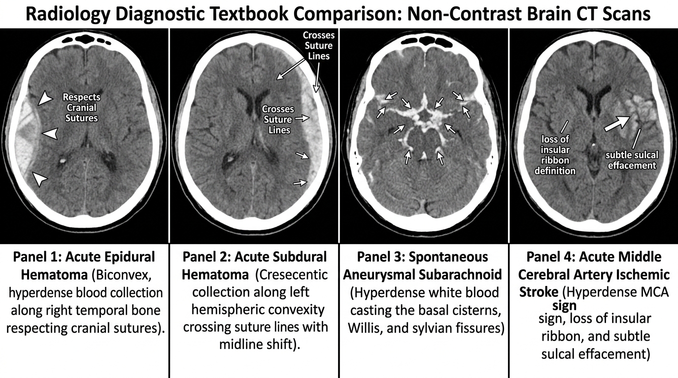
Four-panel non-contrast head CT emergency neuroimaging comparison: (A) Acute epidural hematoma (biconvex lenticular hyperdensity respecting cranial suture lines); (B) Acute subdural hematoma (crescentic hyperdensity crossing suture lines with marked midline shift and sulcal effacement); (C) Aneurysmal subarachnoid hemorrhage (dense hyperattenuating blood casting basal cisterns, sylvian fissures, and interhemispheric fissure); (D) Acute ischemic stroke (early loss of insular ribbon, obscuration of lentiform nucleus, and hyperdense middle cerebral artery sign).
“Non-Contrast Head CT Emergency Neuroimaging Suite” by ClinInterpret Medical Education Editorial Board — CC BY 4.0. View Full Image.
Common Pitfalls & Diagnostic Traps
- Mistaking a subacute isodense subdural hematoma for normal brain; because subacute blood matches brain cortex (+35 HU), look for indirect mass effect: unilateral sulcal effacement, inward displacement of the grey-white junction ('white matter buckling'), or compression of the lateral ventricle.
- Failing to look for hyperdense acute thrombus in cerebral vessels; the 'hyperdense MCA sign' or 'hyperdense basilar artery sign' may be the only visual finding in the first 2 hours of catastrophic ischemic stroke.
- Relying on a normal NCCT to rule out acute ischemic stroke; over 50% of acute ischemic strokes show a completely normal NCCT in the first 3 hours. A normal CT serves primarily to rule out hemorrhage before administering thrombolytics.
- Missing bilateral symmetrical subdural hematomas; because mass effect is equal on both sides, the midline may not be shifted. Look for 'rabbit-ear' compressed, slit-like lateral ventricles.
1Overview and Physiology
Non-Contrast Computed Tomography (NCCT) of the brain is the primary neuroimaging modality for acute neurological emergencies worldwide. It utilizes a rotating X-ray tube and solid-state multidetector array to acquire cross-sectional volumetric slices of the cranium, reconstructing tissue attenuation matrices mapped onto the universal Hounsfield Unit (HU) scale.
2The Physics of Attenuation and Hounsfield Units (HU)
The Hounsfield Unit scale standardizes tissue electron density relative to water and air:
$HU = 1000 × left( frac{µ_tissue - µ_water}{µ_water} right)$
$begin{array}{|l|l|l|} hline textbf{Intracranial Tissue / Fluid} & textbf{Typical Attenuation (HU)} & textbf{Visual Shade} \ hline Air (Sinuses, Mastoid Cells, Pneumocephalus) & -1000 to -800 & Pure Black \ Fat (Lipoma, Dermoid, Teratoma) & -100 to -50 & Dark Grey \ Cerebrospinal Fluid (CSF in Ventricles/Sulci) & 0 to +15 & Dark Grey / Black \ Cytotoxic / Vasogenic Edema & +15 to +25 & Hypodense Grey \ Cerebral White Matter & +25 to +30 & Medium Grey \ Cerebral Grey Matter (Cortex, Basal Ganglia) & +35 to +45 & Lighter Grey \ Clotted Acute Blood (Hemoglobin) & +60 to +90 & Bright Hyperdense White \ Calcification / Pineal / Choroid Plexus & +100 to +300 & Dense White \ Cortical Bone / Skull Table & +700 to +2000+ & Solid Bright White \ hline end{array}$
Grey matter contains higher cell density and protein content than myelin-rich white matter, generating a baseline attenuation difference of 5–10 HU. Preservation of this crisp grey-white matter interface is the hallmark of healthy cerebral parenchyma.
3Technique and Quality Checks
Standard emergency brain CT protocols utilize helical non-contrast volumetric scanning from the skull base (foramen magnum) through the vertex (0.625–1.25 mm slice thickness reconstructed into 3–5 mm slices).
41. Motion and Streak Artifacts
- Patient Motion: Induces concentric arcs and blurring that obscure subtle subdural fluid or early cortical ischemic changes.
- Beam Hardening / Hounsfield Artifacts: Occurs as low-energy X-ray photons are preferentially absorbed by dense bone of the petrous temporal ridges, causing dark streak artifacts across the posterior fossa (brainstem and cerebellum). Thin-slice (1.0 mm) reconstruction and iterative algorithms minimize this distortion.
52. Window Width (W) and Window Level (L) Optimization
Because the human eye can distinguish only approximately 20 to 30 distinct shades of grey, the CT console uses windowing:
- Standard Brain Window ($W: 80, L: 40$): Optimizes the subtle grey-white matter differentiation of brain parenchyma.
- Stroke Window ($W: 30\text{–}40, L: 30\text{–}35$): Narrow window width increases visual contrast, rendering subtle 2–3 HU drops in attenuation (cytotoxic edema) readily visible.
- Subdural Window ($W: 150\text{–}200, L: 50\text{–}75$): Separates dense cortical bone from adjacent hyperdense acute subdural blood.
- Bone Window ($W: 2500\text{–}3000, L: 500\text{–}600$): Suppresses soft tissue completely to inspect for subtle linear, depressed, or skull base fractures.
6Normal Anatomy
71. Ventricular System and Basal Cisterns
- Lateral Ventricles: Symmetrical frontal horns, bodies, and occipital horns; small midline septum pellucidum separates frontal horns.
- Third Ventricle: Slit-like midline cavity flanked by the medial thalami.
- Fourth Ventricle: Midline diamond-shaped structure in the posterior fossa between the pons/medulla and cerebellum.
- Basal Cisterns (The "Safety Valve" of the Brain):
- Suprasellar Cistern: Star-shaped pentagonal or hexagonal CSF space above the sella turcica.
- Ambient and Quadrigeminal Cisterns: CSF encircling the midbrain.
- Prepontine and Premedullary Cisterns: CSF anterior to the brainstem.
- Clinical Significance: Effacement or obliteration of the basal cisterns indicates critically elevated intracranial pressure (ICP) and imminent brainstem herniation.
82. Deep Grey Matter Nuclei
- Caudate Nucleus: Bulges into the lateral wall of the frontal horns of the lateral ventricles.
- Internal Capsule: Dense white matter pathway separated into the anterior limb, genu, and posterior limb (carrying the corticospinal motor tract).
- Lentiform Nucleus: Wedge-shaped deep grey structure comprising the lateral putamen and medial globus pallidus.
- Thalamus: Large egg-shaped sensory relay station flanking the third ventricle.
9Key Signs and Patterns
[ Pathology ]                      [ Radiological Mechanism ]                     [ Radiographic Sign ]
---------------------------------------------------------------------------------------------------------
Middle Meningeal Artery Rupture   --> Bleeding strips periosteum until sutures  --> Biconvex (Lenticular) Hyperdensity
Bridging Cortical Vein Tear       --> Bleeding spreads freely in subdural space --> Crescentic Hemispheric Hyperdensity
Aneurysmal Subarachnoid Rupture   --> Blood fills CSF spaces                     --> Star-shaped Hyperdense Cisterns
Acute MCA Thrombus                --> Dense packed clot in vessel lumen         --> Hyperdense MCA Sign
Early MCA Cytotoxic Edema         --> Sodium/water influx into insular cortex   --> Loss of Insular Ribbon
Middle Fossa Temporal Mass        --> Uncus displaces midbrain posteriorly      --> Kernohan's Notch / Uncal Herniation
Chronic Subdural Re-bleeding --> Fluid levels between lysis and new blood --> Hematocrit / "Swirl" Sign101. Blood Densities by Temporal Age
As extravasated blood ages, hemoglobin undergoes chemical denaturation:
- Hyperacute Blood ($< 3\ \text{hours}$): Non-clotted liquid blood (+45 to +60 HU), occasionally showing the swirl sign (areas of hypodensity representing active, unretracted bleeding).
- Acute Blood ($3\ \text{hours to } 3\text{–}7\ \text{days}$): Clot retraction concentrates hemoglobin to +60 to +90 HU (bright white).
- Subacute Blood ($1\text{ to } 3\ \text{weeks}$): Enzymatic protein breakdown decreases attenuation by roughly 1.5 HU per day, transitioning from isodense (+30 to +45 HU) to dark.
- Chronic Blood ($> 3\ \text{weeks}$): Fully liquefied collection (+0 to +20 HU) resembling water/CSF.
112. The 10-Point ASPECTS Score (Alberta Stroke Programme Early CT Score)
Evaluates middle cerebral artery (MCA) territory early ischemia on two axial CT slices:
- Subcortical (3 points): Caudate (C), Lentiform (L), Internal Capsule (IC).
- Cortical (7 points): Insular cortex (I), M1 (anterior MCA), M2 (MCA lateral to insula), M3 (posterior MCA), M4 (superior MCA anterior), M5 (superior MCA lateral), M6 (superior MCA posterior).
- Score Interpretation: Starts at 10. Subtract 1 point for every region showing hypodensity or loss of grey-white differentiation. An ASPECTS score ≥ 6 qualifies patients for mechanical thrombectomy up to 24 hours under modern clinical guidelines.
12Indications
- Acute Head Trauma: Following Canadian CT Head Rules or New Orleans Criteria (GCS < 15 at 2 hours, suspected open/depressed fracture, signs of basilar skull fracture, ≥ 2 vomiting episodes, age ≥ 65, retrograde amnesia ≥ 30 minutes).
- Acute Stroke Presentation: Immediate exclusion of intracranial hemorrhage prior to IV thrombolytic administration (alteplase / tenecteplase).
- Thunderclap Headache: Exclusion of aneurysmal subarachnoid hemorrhage (sensitivity > 99% within 6 hours of onset).
- Sudden Altered Mental Status / Coma: Assessing acute mass effect, hydrocephalus, or brainstem compression.
- New-Onset Seizure or Sudden Focal Neurological Deficits: Evaluating mass lesions, abscesses, or acute infarcts.
13Systematic Interpretation Steps
Follow the classic mnemonic B-L-O-O-D-S or Blood-Cisterns-Brain-Ventricles-Bone:
[ Step 1: Symmetry & Quality ] --> Verify no motion, inspect grey-white differentiation.
                                          |
                                          v
[ B: Blood ]                   --> Inspect for hyperdensity: Epidural, Subdural,
                                   Subarachnoid, Intracerebral, Intraventricular.
                                          |
                                          v
[ C: Cisterns ]                --> Suprasellar star intact? Ambient cistern patent?
                                   Quadrigeminal space symmetrical?
                                          |
                                          v
[ B: Brain Parenchyma ]        --> Grey-white matter interface crisp? Insular ribbon present?
                                   Lentiform nucleus sharp? Sulci symmetrical?
                                          |
                                          v
[ V: Ventricles ]              --> Symmetrical? Dilated (Hydrocephalus) vs. Effaced?
                                   Midline shift present? (Measure septum pellucidum deviation in mm).
                                          |
                                          v
[ B: Bone ]                    --> Switch to Bone Window: Linear fractures, skull base fracture
                                   (sphenoid sinus fluid level), mastoid opacification.
                                          |
                                          v
[ S: Soft Tissues ] --> Subgaleal hematoma, facial fractures, pneumocephalus.14Abnormal Patterns and Differential Diagnosis
151. Intracranial Herniation Syndromes
- Subfalcine (Cingulate) Herniation: Mass pushes the cingulate gyrus beneath the rigid falx cerebri. Causes compression of the ipsilateral lateral ventricle, contralateral hydrocephalus (foramen of Monro obstruction), and risk of anterior cerebral artery (ACA) infarction.
- Descending Transtentorial (Uncal) Herniation: Medial temporal lobe (uncus) is forced downward over the free tentorial edge. Compresses:
- Ipsilateral Third Cranial Nerve: Dilated, fixed pupil ("blown pupil").
- Ipsilateral Posterior Cerebral Artery (PCA): Occipital lobe infarction (cortical blindness).
- Contralateral Cerebral Peduncle against Kernohan's Notch: Paradoxical ipsilateral hemiparesis.
- Tonsillar Herniation: Cerebellar tonsils are driven downward through the foramen magnum. Compresses the medullary respiratory and cardiac centers, leading to acute respiratory arrest and death.
162. Intracerebral Hemorrhage (ICH) Etiologies
- Hypertensive Microangiopathy: Deep parenchymal hemorrhage in classic locations: Basal ganglia (putamen 50%), Thalamus (15%), Pons (10%), Cerebellum (10%).
- Cerebral Amyloid Angiopathy (CAA): Recurrent, superficial lobar hemorrhages in elderly patients, typically sparing the deep basal ganglia.
- Vascular Malformations (AVM / Cavernoma): Lobar or atypical hemorrhage in younger patients (< 45 years).
17Limitations and Pitfalls
- Normal CT in Early Stroke: An ischemic stroke can appear entirely normal on NCCT during the first 3 to 6 hours. Diffusion-weighted MRI (DWI) is vastly more sensitive (> 95%) in early ischemia.
- Subarachnoid Hemorrhage Beyond 24 Hours: NCCT sensitivity for SAH drops from > 99% within 6 hours to ≈ 85% at 24 hours, and < 50% by 1 week as blood cells lyse. If clinical suspicion remains high and CT is normal after 6 hours, Lumbar Puncture with CSF Spectrophotometry for Xanthochromia is mandatory!
- Posterior Fossa Pathology: Beam hardening artifact from the petrous ridges significantly degrades visualization of the brainstem and cerebellum on CT. MRI is the modality of choice for posterior circulation strokes, vestibular schwannomas, and pontine tumors.
18Critical Findings and Red Flags
[ Midline Shift > 5 mm ]         --> High risk of neurological herniation and clinical deterioration.
                                     ACTION: Urgent neurosurgical consultation, hyperosmolar therapy
                                     (Mannitol 20% or 3% Hypertonic Saline), elevation of head to 30°.

[ Acute Hydrocephalus ]          --> Ballooned temporal horns of the lateral ventricles (> 2 mm width)
                                     with periventricular hypodensity (transependymal CSF resorption).
                                     ACTION: Emergency external ventricular drain (EVD) placement.

[ Tension Pneumocephalus ]       --> "Mount Fuji Sign": Bilateral subdural air compressing the frontal
                                     lobes, creating a widened tented interhemispheric gap.
                                     ACTION: Emergency surgical burr-hole evacuation of trapped air.

[ Basilar Artery Occlusion ]     --> Hyperdense Basilar Artery sign in a comatose or "locked-in" patient.
ACTION: Immediate CTA and emergency mechanical thrombectomy.19Exam Pearls
- The "Mount Fuji Sign": Pathognomonic for tension pneumocephalus, where subdural air separates and compresses both frontal lobes.
- The "Empty Delta Sign": Seen on contrast-enhanced CT head or CT venography; non-enhancing triangular thrombus surrounded by enhancing collateral dural channels in Superior Sagittal Sinus Thrombosis.
- Aspects of the "Swirl Sign": Non-contrast CT swirl sign represents low-attenuation areas of active, unretracted extravasating blood within an acute hyperdense hematoma; highly predictive of hematoma expansion.
- Linear vs Suture Line: Suture lines (coronal, sagittal, lambdoid) have interdigitating, sclerotic, jagged edges and occur in predictable anatomical locations. Traumatic fractures are sharp, lucent, non-sclerotic lines that can cross sutures.
Related Investigations & Companion Tests
Attributed Textbooks & Clinical Guidelines
- Smith AB et al. Brain CT: Neuroimaging in Emergency Medicine and Stroke. Continuum (Minneap Minn). 2021;27(5):1230-1258
- Powers WJ et al. Guidelines for the Early Management of Patients With Acute Ischemic Stroke: 2019 Update. Stroke. 2019;50(12):e344-e418
- Osborn AG. Osborn's Brain: Imaging, Pathology, and Anatomy. 2nd ed. Elsevier, 2017
What is the Hounsfield Unit (HU) scale, and what are the standard densities for CSF, brain parenchyma, acute blood, and bone?
Notes & Highlights
Saved locally onlyA 23-year-old hockey player sustains a forceful blow to the right pterion. He experiences a 2-minute loss of consciousness, followed by a 45-minute 'lucid interval' where he answers orientation questions correctly. Two hours later, he rapidly deteriorates with a GCS drop from 15 to 7, a fixed and dilated right pupil, and left hemiparesis. Urgent NCCT brain reveals a biconvex, high-attenuation extra-axial collection along the right temporoparietal convexity that abuts the coronal suture without crossing it, causing 8 mm of midline shift and compression of the right lateral ventricle. What is the diagnosis and vessel of origin?
A 56-year-old female presents to the emergency department clutching her head, crying out that she has experienced the 'worst headache of my life' (thunderclap headache peaking in under 60 seconds). Neurological examination shows marked nuchal rigidity without focal motor weakness. Blood pressure is 185/105 mmHg. Non-contrast CT of the brain performed 3 hours after onset demonstrates high-attenuation cast-like hyperdensity (+65 HU) filling the basal cisterns, ambient cisterns, and extending into the anterior interhemispheric fissure. What is the diagnosis and next mandatory diagnostic investigation?
A 71-year-old male presents with sudden left-sided hemiplegia and left facial droop starting 90 minutes ago. Baseline blood pressure is 168/94 mmHg, blood glucose is 112 mg/dL. An immediate NCCT brain shows no acute intracranial hemorrhage. However, reviewing the early ischemic stroke parenchyma reveals blurring of the right insular cortex margin compared to the normal left insula, and slight hypodensity with swelling of the right putamen (obscuration of the lentiform nucleus). The total ASPECTS score is calculated at 8. What is the clinical interpretation and management?