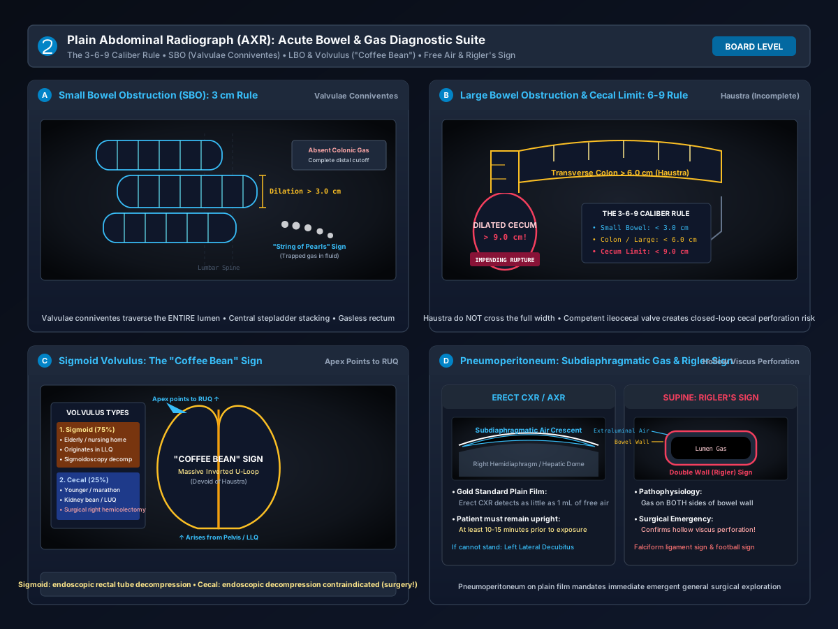
Four-panel plain abdominal radiography diagnostic suite: (A) Small bowel obstruction showing central stacked loops >3 cm with complete valvulae conniventes and 'string of pearls' sign; (B) Large bowel obstruction showing peripheral colonic frame >6 cm with incomplete haustra and cecal distension limit (>9 cm); (C) Sigmoid volvulus demonstrating classic 'coffee bean' sign arising from LLQ with apex pointing to RUQ; (D) Pneumoperitoneum demonstrating erect subdiaphragmatic gas crescent and supine Rigler double-wall sign.
“Abdominal Radiography: SBO vs LBO, 3-6-9 Rule, Volvulus, and Rigler's Sign” by ClinInterpret Medical Education Editorial Board — CC BY 4.0. View Full Image.
Common Pitfalls & Diagnostic Traps
- Diagnosing small bowel obstruction on a supine film when the bowel caliber is 2.5 cm; always remember the 3-6-9 rule (small bowel must exceed 3.0 cm, colon 6.0 cm, cecum 9.0 cm).
- Confusing the transverse colon for small bowel; trace the folds to confirm whether they cross the lumen completely (valvulae conniventes in small bowel) or terminate partially (haustra in colon).
- Attempting endoscopic detorsion on a Cecal Volvulus; endoscopic reduction is successful only in Sigmoid Volvulus. In Cecal Volvulus, colonoscopy carries high perforation risk and surgery is mandatory.
- Dismissing free air because an erect CXR was acquired immediately after standing; patients must remain erect for 10 to 15 minutes before exposure to permit low-density gas to migrate beneath the diaphragm.
1Overview and Physiology
Plain Abdominal Radiography—frequently termed the Kidney-Ureter-Bladder (KUB) or plain abdominal film (AXR)—is a rapid, widely available projectional imaging examination utilizing ionizing radiation to survey the distribution of abdominal gas, soft tissue organ silhouettes, and pathological calcifications.
2Physical Radiodensity and Gas Distribution
In the healthy abdomen, the distribution of gas is strictly physiological:
- Stomach: Almost always contains a variable volume of gas forming the gastric air bubble (fundus).
- Small Bowel: Typically contains minimal gas (<2 to 3 small non-dilated loops with calibers <2.5 cm) because swallowed atmospheric air is rapidly propelled or absorbed.
- Colon & Rectum: Contains variable gas and mottled, granular particulate material representing stool, predominantly in the cecum, ascending colon, and rectum.
When mechanical transit is obstructed, proximal peristaltic contractions hyper-propel fluid and gas against the transition point. Secretions accumulate (saliva, gastric juice, bile, pancreatic fluid: ~7--9 liters/day), resulting in bowel wall stretch, third-space fluid sequestration, venous congestion, and progressive intraluminal distension.
3Technique and Quality Checks
- Standard Projections:
- Supine AP Abdominal Radiograph: The standard baseline view. Demonstrates bowel loop calibers, anatomical distribution, mass effect, calcifications, and the supine signs of free air (Rigler's sign).
- Erect Chest Radiograph (PA CXR): The single most sensitive plain radiographic examination for detecting pneumoperitoneum (can detect as little as 1 mL of free subdiaphragmatic air).
- Left Lateral Decubitus Abdomen: Acquired when the patient is physically unable to sit or stand for an erect chest film. The patient lies left-side-down for 10 minutes; free peritoneal air rises and layers along the lateral liver edge and right flank peritoneal gutter, where it cannot be confused with the gastric bubble.
- Field of View Verification:
- The radiograph must extend superiorly from the diaphragmatic domes down to the inferior pubic symphysis to ensure the hernial orifices (inguinal and femoral canals) and pelvis are fully visualized.
- Exposure and Penetration:
- Under-penetration fails to visualize renal and psoas margins; over-penetration burns out faint extraluminal gas bubbles.
4Normal Anatomy
Systematic appraisal begins with recognition of baseline normal anatomical structures:
- The Gastrointestinal Tract:
- Small Bowel: Located centrally. Mucosal folds (Valvulae Conniventes / Plicae Circulares) are closely spaced (1--2 mm) and cross the entire diameter of the bowel lumen from wall to wall.
- Large Bowel (Colon): Located peripherally forming an inverted U-frame. Features Haustra (folds separated by plicae semilunares) that extend only partially (one-third to two-thirds) across the luminal width.
- The 3-6-9 Caliber Rule:
- Small Bowel: Maximum normal luminal diameter is $< 3.0\ \text{cm}$.
- Colon (Large Bowel): Maximum normal luminal diameter is $< 6.0\ \text{cm}$.
- Cecum: Maximum normal luminal diameter is $< 9.0\ \text{cm}$.
- Retroperitoneal Soft Tissue Silhouettes:
- Distinct linear fat planes outlining the bilateral psoas muscle borders (loss of psoas stripe suggests retroperitoneal fluid, hematoma, or abscess).
- Outlines of the liver, spleen, and bilateral renal silhouettes.
5Key Signs and Patterns
$begin{array}{|l|l|l|} hline textbf{Radiological Sign} & textbf{Visual Morphological Appearance} & textbf{Pathological Significance} \ hline textbf{Valvulae Conniventes} & Thin, parallel radiodense lines spanning full lumen & text{Confirms Small Bowel dilation (>3\ \text{cm})} \ textbf{Haustra} & Thick, widely spaced sacculations spanning partial lumen & text{Confirms Colonic / Large Bowel dilation (>6\ \text{cm})} \ textbf{String of Pearls Sign} & Row of tiny, rounded gas bubbles trapped in valvulae & High-grade mechanical Small Bowel Obstruction (SBO) \ textbf{Stepladder Sign} & Vertically stacked horizontal air-fluid levels on erect film & Mechanical small bowel transit blockage \ textbf{Coffee Bean Sign} & Massive inverted U-loop from LLQ to RUQ with central seam & Sigmoid Volvulus (pathognomonic) \ textbf{Rigler's Sign} & Both inner (mucosal) and outer (serosal) bowel walls visible & Pneumoperitoneum (free peritoneal air on supine film) \ textbf{Falciform Ligament Sign} & Vertical linear opacity outlined by gas in RUQ & Extensive free air surrounding ligamentum teres \ textbf{Football Sign} & Colossal, oval lucency outlining entire peritoneal cavity & Massive pneumoperitoneum (most common in neonates) \ hline end{array}$
6Indications
- Suspected Bowel Obstruction: Nausea, bilious vomiting, abdominal distension, high-pitched hyperactive or absent bowel sounds, and obstipation.
- Evaluation for Hollow Viscus Perforation: Sudden onset severe abdominal pain, peritonitis, board-like rigidity (erect CXR / decubitus view).
- Assessment of Known Colonic Volvulus: Monitoring detorsion response or acute recurrence in institutionalized patients.
- Localization of Radiopaque Foreign Bodies or Interventional Lines: Confirming nasogastric (NG) tube tip placement in the gastric fundus/antrum past the gastroesophageal junction, or locating swallowed radiopaque objects.
- Surveillance of Toxic Megacolon: Serial caliber measurements in acute severe ulcerative colitis or C. difficile pseudomembranous colitis.
7Systematic Interpretation Steps
Step 1: Check Quality & Projection
   ├─ Confirm supine AP vs erect CXR vs left lateral decubitus
   └─ Ensure coverage from diaphragmatic domes down to pubic symphysis

Step 2: Inspect for Free Extraluminal Air (Pneumoperitoneum)
   ├─ Look under the right hemidiaphragm for subdiaphragmatic gas crescents
   ├─ Inspect for Rigler's Double Wall Sign across central bowel loops
   └─ Search for the falciform ligament sign and football sign

Step 3: Analyze Bowel Caliber (The 3-6-9 Rule)
   ├─ Are small bowel loops > 3.0 cm? (Central, valvulae conniventes wall-to-wall)
   ├─ Is the transverse colon > 6.0 cm? (Peripheral, haustra partial folds)
   └─ Is the cecum dilated >= 9.0 cm? (Impending closed-loop rupture!)

Step 4: Differentiate SBO vs LBO vs Volvulus
   ├─ SBO: Centrally stacked dilated loops + empty distal colon/rectum
   ├─ LBO: Dilated colonic frame up to transition point + variable cecal dilation
   └─ Volvulus: Giant 'Coffee Bean' loop (Sigmoid) or teardrop loop (Cecal)

Step 5: Survey Bones, Calcifications, and Soft Tissue Stripes
   ├─ Inspect psoas muscle contours and properitoneal flank fat stripes
   ├─ Check for renal calculi, gallstones, or vascular aortic calcifications
└─ Inspect pelvic bones and spine for lytic/blastic lesions or fractures8Abnormal Patterns and Differential Diagnosis
91. Mechanical Small Bowel Obstruction (SBO)
- Radiological Findings: Multiple centrally located, dilated loops (>3 cm) displaying transverse mucosal lines (valvulae conniventes). On erect films, multiple air-fluid levels in a 'stepladder' distribution or 'string of pearls' sign. The colon and rectum are decompressed and gasless.
- Differential Diagnosis: Adynamic paralytic ileus (gas uniformly present in both small bowel and colon without cutoff), gastroenteritis, mesenteric ischemia.
102. Large Bowel Obstruction (LBO)
- Radiological Findings: Continuous colonic distension (>6 cm) extending from the cecum along the ascending, transverse, and descending colon to the anatomical point of obstruction (e.g. colorectal adenocarcinoma, diverticular stricture).
- The Role of the Ileocecal Valve:
- Competent Valve (Closed Loop): Colonic gas cannot reflux retrograde into the small bowel; massive cecal distension ensues (≥ 9--12 cm), carrying extremely high perforation risk.
- Incompetent Valve: Colonic gas refluxes into the ileum, decompressing the cecum and mimicking distal SBO.
113. Sigmoid vs Cecal Volvulus
- Sigmoid Volvulus (75%): Arises from the pelvis / left lower quadrant and extends into the right upper quadrant as a giant, smooth-walled loop resembling a Coffee Bean. The bowel is devoid of haustra. The central crease represents the apposed inner walls.
- Cecal Volvulus (25%): Arises from mobile cecal mesentery in the right lower quadrant, twisting and displacing a comma-shaped or teardrop gas collection into the left upper quadrant or epigastrium.
12Limitations and Pitfalls
- Diagnostic Superiority of Contrast CT: Plain abdominal radiographs have an overall sensitivity of only $50\text{--}70\%$ for small bowel obstruction and cannot reliably identify bowel ischemia, strangulation, closed loops, internal hernias, or transition point etiology. Contrast-enhanced CT of the abdomen and pelvis is the definitive gold standard.
- The Fluid-Filled Closed Loop Pitfall: Completely fluid-filled obstructed bowel loops contain zero intraluminal gas, rendering them invisible on plain radiographs ('pseudotumor' or gasless abdomen), falsely reassuring the clinician.
- Chilaiditi Sign vs Free Air: Hepatodiaphragmatic interposition of the colon (Chilaiditi sign) mimics subdiaphragmatic free air. Look closely for colonic haustral folds within the gas collection to avoid unnecessary emergency laparotomy!
13Critical Findings and Red Flags
> [!CAUTION] > Emergency Surgical Escalation Triggers: > 1. Subdiaphragmatic Free Gas or Rigler's Sign: Establishes gastrointestinal hollow viscus perforation; requires urgent surgical consultation and immediate intravenous broad-spectrum antibiotics. > 2. Cecal Dilation $\ge 9\text{--}12\ \text{cm}$: Laplace's Law dictates that wall tension is proportional to diameter (T = P × r); a cecum >9 cm has the thinnest wall and highest risk of ischemic blowout perforation. > 3. Pneumatosis Intestinalis and Portal Venous Gas: Thin linear gas ribbons within the bowel wall combined with branching peripheral liver gas indicates transmural bowel necrosis and infarction.
14Exam Pearls
- Laplace's Law and the Cecum: Because the cecum has the largest resting diameter of the large bowel, wall tension (T = P × r) is highest in the cecum for any given intraluminal pressure. Thus, in distal colonic obstruction, the cecum always perforates first!
- Rigler's Sign Mechanism: Normally, the bowel wall appears as a single soft-tissue density because the outer serosa abuts peritoneal fluid. When free peritoneal gas surrounds the outside, gas is present on both sides, transforming the bowel wall into a distinct double radiopaque line.
- The Detorsion Management Pearl: Sigmoid volvulus without peritonitis is initially treated with endoscopic sigmoidoscopic decompression and rectal tube placement. Cecal volvulus cannot be decompressed endoscopically and requires immediate operative surgery (right hemicolectomy).
Related Investigations & Companion Tests
Attributed Textbooks & Clinical Guidelines
- Herring W. Learning Radiology: Recognizing the Basics. 4th ed. Elsevier, 2019
- Silva AC et al. Bowel Obstruction: What the Radiologist Needs to Know. Radiographics. 2009;29(2):423-439
- Feldman M et al. Sleisenger and Fordtran's Gastrointestinal and Liver Disease. 11th ed. Elsevier, 2021
- Grainger & Allison's Diagnostic Radiology. 7th ed. Elsevier, 2021
What is the classic '3-6-9 Caliber Rule' for maximum normal bowel diameters on plain abdominal radiography?
Notes & Highlights
Saved locally onlyA 62-year-old female with a prior history of open hysterectomy presents to the emergency department with cramping mid-abdominal pain, bilious vomiting, and obstipation for 24 hours. A supine abdominal radiograph demonstrates centrally stacked, dilated loops of bowel measuring 4.2 cm in caliber. Thin, linear radiodense bands extend completely across the lumen from one wall to the opposite wall. The ascending colon, transverse colon, and rectum contain zero gas. What is the diagnosis and anatomical hallmark?
An 82-year-old male resident of a nursing home with chronic constipation and Parkinson's disease presents with severe abdominal distension and obstipation. An abdominal radiograph reveals a colossal, smooth-walled gas-filled loop devoid of haustra arising from the pelvis and left lower quadrant, extending cranially up to the T8 vertebral level with its apex directed into the right hypochondrium. A dense central cleft separates the two dilated apposed limbs. Distal rectal gas is absent. What is the diagnosis and initial management?
A 48-year-old male with a history of untreated peptic ulcer disease presents to the emergency department with sudden, catastrophic 'thunderclap' epigastric pain and board-like abdominal rigidity. The patient is too weak to sit or stand for an upright chest radiograph. A portable supine abdominal radiograph is acquired. On the supine view, both the mucosal inner lining and the outer serosal margin of several loops of small bowel in the mid-abdomen are sharply visualized as distinct radiopaque lines outlined by radiolucent gas. What is this sign, and what pathology does it establish?