Rise/fall curves for troponin vs CK-MB vs myoglobin after infarction.
Schematic illustration, not a patient tracing.
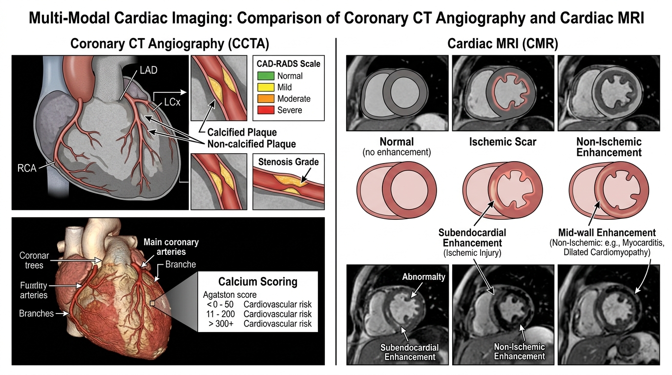
Multi-modal cardiac imaging diagnostic framework. Left: Coronary CT angiography showing calcium scoring, plaque morphology, and CAD-RADS stenosis grading. Right: Cardiac MRI short-axis inversion recovery slices illustrating ischemic subendocardial scar versus non-ischemic mid-wall enhancement.
“Multi-Modal Cardiac Imaging: CCTA and CMR Diagnostic Patterns” by ClinInterpret Medical Education Editorial Board — CC BY 4.0. View Full Image.
1Overview and Physiology
Multi-modal cardiac imaging—specifically Coronary Computed Tomography Angiography (CCTA), Coronary Artery Calcium (CAC) Scoring, and Cardiac Magnetic Resonance (CMR)—represents the pinnacle of non-invasive cardiovascular diagnostics. Where plain radiographs and standard surface electrocardiography provide indirect or functional clues, advanced cross-sectional cardiac imaging delivers direct anatomical, histological, and tissue-characterization resolution of the coronary tree and myocardium.
Coronary CT capitalizes on rapid slip-ring gantry rotation, sub-millimeter multi-detector arrays, and prospectively triggered ECG gating to capture motion-free images of the coronary lumen, calcified and non-calcified atherosclerotic plaques, and congenital coronary anomalies during mid-diastole.
Conversely, Cardiac Magnetic Resonance (CMR) utilizes intrinsic hydrogen proton magnetic properties under static and radiofrequency magnetic fields. CMR provides the clinical reference standard for biventricular volumes, ejection fractions, tissue edema, parametric T1/T2 mapping, and Late Gadolinium Enhancement (LGE)—the definitive technique for differentiating ischemic myocardial scar from non-ischemic cardiomyopathies, active myocarditis, and infiltrative storage disorders.
2Technique and Quality Checks
31. Coronary CT Angiography & Calcium Scoring Preparation
- Heart Rate Optimization: Target resting heart rate <60 bpm (ideally 50--55 bpm) using oral beta-blockers (e.g., metoprolol tartrate 50--100 mg 1 hour prior or IV metoprolol 5 mg increments intra-procedural) or ivabradine. Heart rate variability and arrhythmias (atrial fibrillation) degrade diagnostic lumen interpretability.
- Sublingual Vasodilation: Sublingual nitroglycerin (0.4--0.8 mg spray or tablet) administered 3–5 minutes before contrast injection maximizes epicardial coronary caliber and diagnostic visualization of distal branches.
- Contrast Dynamics: High-iodine-concentration contrast (350--400 mg I/mL) administered at 5--7 mL/s followed by a saline chaser. Bolus tracking in the ascending aorta triggers acquisition at peak coronary enhancement (>300 HU).
- Radiation Optimization: Prospective ECG triggering (axial step-and-shoot) limits exposure to a narrow diastolic window (70--80% of R-R interval), reducing radiation exposure to <1--3 mSv compared to retrospective spiral acquisition.
42. Cardiac MRI Protocols & Sequences
- Cine Balanced Steady-State Free Precession (b-SSFP): Acquired in standard cardiac planes (long-axis 2-chamber, 3-chamber, 4-chamber, and short-axis ventricular stack) to calculate end-diastolic volume (EDV), end-systolic volume (ESV), stroke volume (SV), and ejection fraction (LVEF/RVEF).
- Late Gadolinium Enhancement (LGE): Inversion-recovery T1-weighted gradient echo sequences acquired 10–15 minutes following intravenous administration of 0.1--0.2 mmol/kg extracellular gadolinium-based contrast agent (GBCA).
- Inversion Time (TI) Optimization: Accurate selection of TI (typically 250--350 ms, confirmed via a Look-Locker scout sequence) to completely null the signal of healthy, uninjured myocardium, making scarred or fibrotic areas appear hyperintense ("bright white").
5Normal Anatomy
Systematic interpretation requires evaluating distinct cardiac compartments:
- Coronary Arterial Tree (CCTA):
- Left Main Coronary Artery (LMCA): Arises from the left coronary sinus, courses between the pulmonary trunk and left atrial appendage, and bifurcates into the Left Anterior Descending (LAD) and Left Circumflex (LCx) arteries.
- Left Anterior Descending (LAD): Courses in the anterior interventricular groove, supplying the anterior wall, anterior septum, and cardiac apex via diagonal branches and septal perforators.
- Left Circumflex (LCx): Travels in the left atrioventricular groove, supplying the lateral and posterolateral left ventricular walls via obtuse marginal (OM) branches.
- Right Coronary Artery (RCA): Originates from the right coronary sinus, courses in the right AV groove, giving rise to acute marginal branches and, in right-dominant circulation (85% of individuals), terminating as the Posterior Descending Artery (PDA) and Posterolateral (PLV) branch.
- Myocardial Architecture (CMR):
- The left ventricle is divided into 17 standardized myocardial segments according to the American Heart Association (AHA) consensus model (basal 6 segments, mid-cavity 6 segments, apical 4 segments, and the true apex).
- Compact myocardium measures 6--10 mm at end-diastole; normal right ventricular free wall measures ≤ 3--4 mm.
6Key Signs and Patterns
71. Agatston Calcium Score Categories
Calculated by multiplying the area of each calcified lesion (>130 HU) by a peak attenuation cofactor:
- Agatston 0: Zero detectable coronary calcium. Associated with extremely low 5-year cardiovascular event rates (<0.1%/year), often termed the "power of zero."
- Agatston 1–99: Mild plaque burden. Early subclinical coronary atherosclerosis; indicates lifestyle intervention and initiation of moderate-intensity statin.
- Agatston 100–299: Moderate atherosclerotic burden. Definite coronary disease; guideline-directed medical therapy with high-intensity statin and aspirin indicated.
- Agatston $\ge 300$ (or $\ge 75^{\text{th}}$ percentile): Severe atherosclerotic disease burden. High annual MACE rate; warrants aggressive primary prevention and consideration of functional ischemia evaluation.
82. CAD-RADS 2.0 Stenosis Grading & High-Risk Plaque Features
- CAD-RADS 0: 0% stenosis (Normal; no plaque).
- CAD-RADS 1: 1--24% stenosis (Minimal stenosis; non-obstructive).
- CAD-RADS 2: 25--49% stenosis (Mild stenosis; non-obstructive).
- CAD-RADS 3: 50--69% stenosis (Moderate stenosis; functional evaluation like FFR-CT or stress imaging recommended).
- CAD-RADS 4A: 70--99% stenosis in 1 or 2 vessels (Severe stenosis; invasive coronary angiography recommended).
- CAD-RADS 4B: ≥ 50% Left Main stenosis or 3-vessel disease with ≥ 70% stenosis (High-risk anatomical CAD; urgent invasive coronary angiography indicated).
- CAD-RADS 5: 100% total occlusion.
- High-Risk Plaque (HRP) Modifiers:
- Low Attenuation Plaque: Core attenuation <30 HU reflecting a necrotic lipid core.
- Positive Remodeling: Outer vessel diameter at lesion >1.1× reference segment.
- Spotty Calcification: Punctate calcium flecks <3 mm within soft plaque.
- Napkin-Ring Sign: Central low-attenuation necrotic core surrounded by a rim of higher attenuation.
93. CMR Late Gadolinium Enhancement (LGE) Pathological Patterns
- Subendocardial to Transmural (Ischemic Pattern):
- Originates at the subendocardium and progresses outward toward the epicardium strictly within the distribution of a single coronary artery (LAD, LCx, or RCA).
- Transmurality <50% predicts functional contractile recovery following revascularization; transmurality >50--75% indicates permanent non-viable scar.
- Mid-Wall / Striae (Non-Ischemic Pattern):
- Sparing of the subendocardium, non-coronary distribution. Characteristic of idiopathic dilated cardiomyopathy (DCM), genetic lamin A/C mutation, and chronic myocarditis.
- Subepicardial / Patchy Pattern:
- Predominantly localized to the inferolateral left ventricular wall. Classic for acute or healed viral myocarditis and systemic sarcoidosis.
- Right Ventricular Insertion Point Enhancement:
- Focal patchy enhancement at the anterior and posterior hinge points where the right ventricle joins the septum. Characteristic of Hypertrophic Cardiomyopathy (HCM) and chronic severe pulmonary arterial hypertension.
- Global Diffuse Subendocardial Non-Coronary LGE:
- Diffuse circumferential subendocardial enhancement with difficulty achieving myocardial nulling; pathognomonic for Cardiac Amyloidosis.
10Indications
- Stable Chest Pain / Angina with Intermediate Pre-Test Probability: CCTA is the Class I recommended first-line anatomical test to rule out obstructive CAD (NICE CG95, ESC Guidelines).
- Cardiovascular Risk Reclassification in Asymptomatic Patients: Coronary Calcium Scoring is indicated in borderline or intermediate-risk patients (ACC/AHA Class IIa) to guide statin decisions.
- Assessment of Myocardial Viability: CMR LGE provides the definitive clinical test to determine whether dysfunctional myocardium will recover after CABG or PCI.
- Differentiation of Unexplained Cardiomyopathies: Investigating new-onset heart failure, non-ischemic LV dysfunction, unexplained ventricular arrhythmias, and suspected cardiac amyloidosis or sarcoidosis.
- Suspected Acute or Subacute Myocarditis: Documenting myocardial edema (T2 STIR) and subepicardial necrosis (LGE) according to updated Lake Louise criteria.
11Systematic Interpretation Steps
Step 1: Technical & Artifact Audit
   ├─ CCTA: Confirm motion-free acquisition, heart rate <60 bpm, contrast attenuation >300 HU
   └─ CMR: Confirm myocardial nulling on Look-Locker scout and absence of respiratory ghosting

Step 2: Quantitative Chamber Assessment (CMR)
   ├─ Measure LV End-Diastolic Volume (EDV), End-Systolic Volume (ESV), and Ejection Fraction
   ├─ Calculate Left Ventricular Mass and end-diastolic wall thickness (normal: 6-10 mm)
   └─ Assess RV dimensions, TAPSE, and RV ejection fraction

Step 3: Coronary Tree Systematic Interrogation (CCTA)
   ├─ Trace LMCA, LAD (including diagonals & septals), LCx (OMs), and RCA (PDA/PLV)
   ├─ Calculate Agatston Calcium Score across all coronary branches
   ├─ Grade maximal luminal diameter stenosis according to CAD-RADS 2.0 (0 to 5)
   └─ Search for high-risk plaque features (low attenuation <30 HU, positive remodeling, napkin-ring)

Step 4: Tissue Characterization & LGE Analysis (CMR)
   ├─ T2 STIR / T2 mapping: Evaluate for active myocardial edema (T2 signal ratio >2.0)
   ├─ Native T1 & ECV mapping: Quantify diffuse extracellular expansion (amyloid, fibrosis)
   └─ LGE Inversion Recovery: Classify enhancement distribution
         ├─ Subendocardial/Transmural (coronary distribution) → Ischemic infarction
         ├─ Mid-wall linear → Dilated cardiomyopathy, myocarditis
         ├─ Subepicardial inferolateral → Acute/post-viral myocarditis, sarcoidosis
         ├─ RV Insertion points → Hypertrophic cardiomyopathy, RV pressure overload
└─ Global diffuse subendocardial → Cardiac amyloidosis12Abnormal Patterns and Differential Diagnosis
| Imaging Finding | Diagnostic Modality | Primary Differential Diagnosis | Confirmatory / Next Actions |
|---|---|---|---|
| Subendocardial LGE in LAD territory (<50% thickness) | Cardiac MRI | Viable ischemic myocardial hibernation | Revascularization (PCI / CABG) indicates high likelihood of functional recovery |
| Transmural LGE in RCA territory (>75% thickness) | Cardiac MRI | Non-viable completed transmural inferior myocardial infarction | Medical therapy; revascularization unlikely to restore contractility in that segment |
| Patchy inferolateral subepicardial LGE + T2 hyperintensity | Cardiac MRI | Acute Viral Myocarditis (Lake Louise criteria positive) | Serum troponin, viral serology, exercise restriction, neurohormonal blockade |
| Mid-wall linear striae LGE in basal interventricular septum | Cardiac MRI | Dilated Cardiomyopathy (DCM) / Lamin A/C cardiomyopathy | Genetic testing, electrophysiological evaluation for sudden cardiac death / ICD |
| Diffuse global subendocardial LGE + failure to null | Cardiac MRI | Cardiac Amyloidosis (AL or ATTR) | Serum free light chains, urine immunofixation, 99mTc-PYP bone scintigraphy |
| CAD-RADS 4B (Left Main $\ge 50\%$ or 3-vessel $\ge 70\%$) | Coronary CT | Anatomically high-risk obstructive coronary artery disease | Urgent invasive coronary angiography and Heart Team evaluation for CABG/PCI |
| CAD-RADS 3 with low-attenuation plaque & napkin-ring sign | Coronary CT | Vulnerable moderate coronary stenosis | High-intensity statin, aspirin, functional FFR-CT or stress perfusion imaging |
13Limitations and Pitfalls
- Heavy Coronary Calcification & "Blooming" Artifact on CT: Dense calcium deposits (Agatston >400--1000) create partial-volume volume-blooming artifacts that artificially exaggerate lumen narrowing, leading to false-positive overestimations of stenosis.
- High Heart Rate and Arrhythmias: Irregular R-R intervals (atrial fibrillation) or tachycardia >65 bpm cause severe phase-misregistration and staircase motion artifacts on CT reconstructions.
- Severe Renal Impairment (eGFR $<30\text{ mL/min/1.73 m}^2$): Relative contraindication to iodinated radiocontrast (Contrast-Induced Nephropathy / PC-AKI) on CT and linear gadolinium agents on MRI due to Nephrogenic Systemic Fibrosis (NSF) risk (macrocyclic agents carry near-zero NSF risk).
- Cardiac Devices & Metallic Artifacts: While most modern pacemakers and ICDs are MR-conditional, generator susceptibility artifacts create signal void and distortion, particularly in the anterior and lateral LV walls.
- Inaccurate Inversion Time (TI) on CMR: Selecting an incorrect inversion time causes incomplete myocardial nulling, creating pseudo-enhancement that mimics subendocardial or patchy scar.
14Critical Findings and Red Flags
- CAD-RADS 4B or CAD-RADS 5: Left main stenosis ≥ 50% or severe proximal multi-vessel disease requires immediate notification of the treating cardiologist and urgent invasive catheterization.
- Coronary Artery Ectopic Origin with Interarterial Course: Malignant anomalous coronary origin (e.g., anomalous left coronary artery arising from the right sinus and coursing between the aorta and pulmonary artery) carries high risk for exercise-induced sudden cardiac death.
- Extensive Myocardial LGE in HCM ($\ge 15\%$ of LV Mass): A major independent risk factor for ventricular fibrillation and sudden cardiac death, serving as a guideline recommendation for primary prevention implantable cardioverter-defibrillator (ICD) placement.
- Left Ventricular Thrombus: Identified on early post-contrast CMR sequences as an avascular non-enhancing intracavitary mass abutting an akinetic scarred segment; warrants immediate systemic anticoagulation.
15Exam Pearls
- Agatston "Power of Zero": In patients with intermediate 10-year ASCVD risk where the decision to start statin therapy is uncertain, a CAC score of 0 can reclassify the patient downward, allowing statin therapy to be safely withheld.
- Ischemic vs Non-Ischemic Golden Rule: Ischemic injury ALWAYS involves the subendocardium because it is the watershed zone most vulnerable to hypoperfusion. Any enhancement that completely spares the subendocardium (e.g., mid-wall or subepicardial) is non-ischemic by definition.
- Viability Cutoff ($50\%$): On board exams, remember the 50% transmural scar threshold on CMR LGE. <50% transmural thickness = viable myocardium that will regain contractility post-bypass; >50% transmural thickness = completed fibrous scar that will not recover.
- Napkin-Ring Sign: Represents the most specific CT imaging hallmark of a high-risk rupture-prone coronary plaque with a large lipid-rich necrotic core.
- Amyloidosis Myocardial Nulling: In cardiac amyloidosis, gadolinium kinetics are altered by massive extracellular interstitial infiltration; the blood pool and myocardium null at virtually identical inversion times, causing the radiologist to struggle finding a normal nulling point.
Related Investigations & Companion Tests
Attributed Textbooks & Clinical Guidelines
- Schulz-Menger J, et al. Standardized image interpretation and post-processing in cardiovascular magnetic resonance: Society for Cardiovascular Magnetic Resonance (SCMR) Board of Trustees Task Force on Standardized Post-Processing. J Cardiovasc Magn Reson. 2020.
- Cury RC, et al. CAD-RADS 2.0: 2022 Coronary Artery Disease - Reporting and Data System: An Expert Consensus Document of SCCT, ACC, ACR, and NASCI. J Am Coll Cardiol. 2022.
- Braunwald's Heart Disease: A Textbook of Cardiovascular Medicine, 12th Edition. Elsevier, 2022.
What are the Agatston Coronary Artery Calcium (CAC) score risk categories and clinical decision thresholds?
Notes & Highlights
Saved locally onlyA 54-year-old male with hypertension and a family history of premature CAD presents for cardiovascular risk evaluation. He is asymptomatic. Non-contrast cardiac CT demonstrates an Agatston Calcium Score of 385 (placing him in the 92nd percentile for age and sex). What is the most appropriate next step in clinical management?
A 32-year-old female presents with atypical chest pain and elevated high-sensitivity troponin 2 weeks following a viral respiratory illness. Coronary angiography shows smooth, normal epicardial coronaries. Cardiac MRI reveals normal left ventricular volumes, preserved ejection fraction (56%), and patchy subepicardial and mid-wall late gadolinium enhancement in the basal-to-mid inferolateral left ventricular wall. What is the most likely diagnosis?
A 66-year-old female with prior anterior myocardial infarction and heart failure (LVEF 30%) is evaluated for surgical revascularization. Cardiac MRI reveals akinesis of the anterior wall and apex. LGE demonstrates hyperenhancement involving 15% of the myocardial wall thickness in the anterior segments. What does this late gadolinium finding indicate regarding myocardial viability?